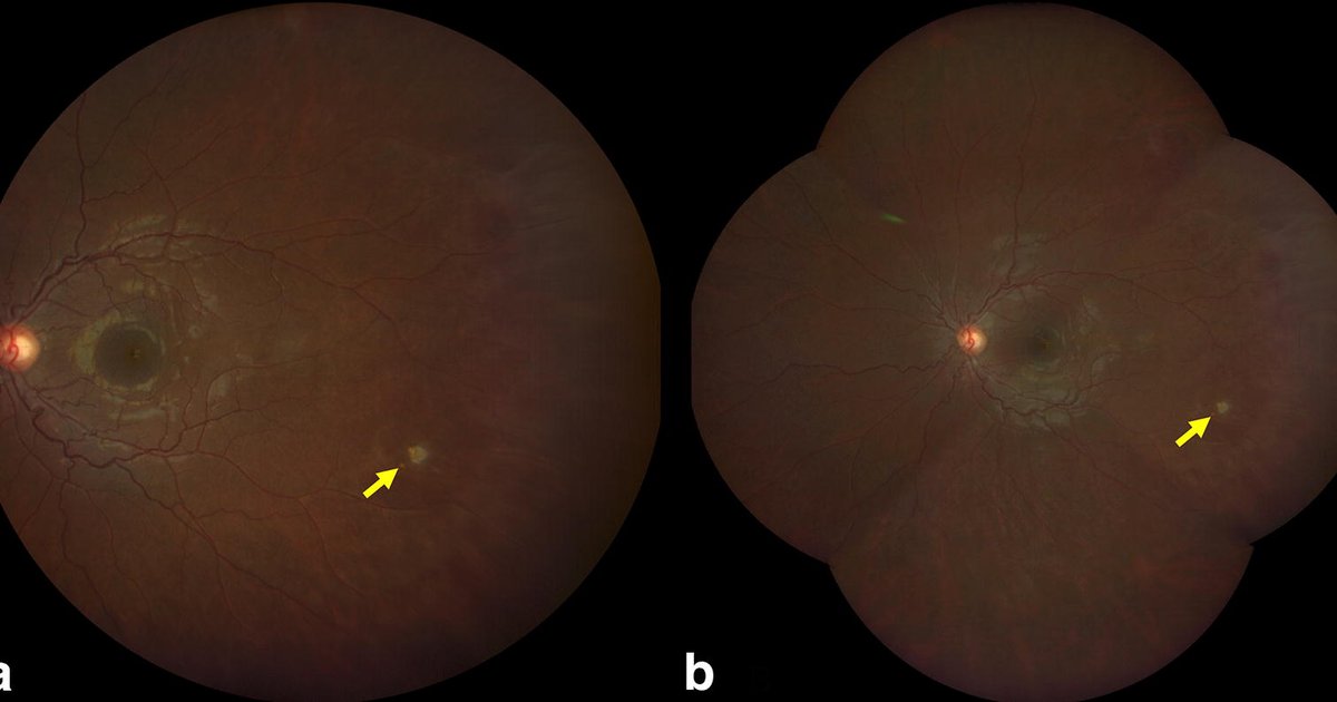
Sickle cell retinopathy is damage to the retina, the light-sensing layer at the back of the eye, caused by sickled red cells blocking its tiny blood vessels. It usually causes no symptoms until it is advanced, which is why the national expert panel recommends that everyone with sickle cell disease see an eye doctor for a dilated eye exam starting at age 10, and every 1 to 2 years after a normal exam (NHLBI Expert Panel, 2014). The risk is highest in hemoglobin SC disease, not only in sickle cell anemia (SS) (Downes et al., 2005). Caught early, the dangerous form can be treated with laser. Sudden floaters, flashes or a shadow over your vision need an eye doctor the same day.
What sickle cell retinopathy is
Sickled cells clog the small arteries at the outer edge of the retina, and the starved tissue sends out signals that grow fragile new vessels. The expert panel describes five stages (NHLBI Expert Panel):
- Stages I and II: blocked arteries at the edge of the retina and rewiring of the vessels around them.
- Stage III: new, fragile vessels grow in a fan shape ("sea fans"). This is proliferative sickle retinopathy, the stage that threatens vision.
- Stage IV: those vessels bleed into the gel that fills the eye (vitreous hemorrhage).
- Stage V: scar tissue pulls the retina away from the back of the eye (retinal detachment).
Unlike diabetic retinopathy, which starts near the center, sickle retinopathy starts at the far edge of the retina, so it can be advanced before you notice anything (AAO EyeWiki). For the diabetic kind, see our guide to diabetic retinopathy in Black adults.
Who is most at risk
The expert panel says everyone with sickle cell disease, and especially people with HbSC, is at risk (NHLBI Expert Panel). The clearest numbers come from a 20-year study in Jamaica that followed children identified at birth. By ages 24 to 26, proliferative retinopathy had developed in 43% of people with HbSC disease and 14% of people with HbSS disease, and new cases arose about five times as often in HbSC (2.5 against 0.5 per 100 people a year) (Downes et al.). The panel notes the peak in HbSC comes at about ages 15 to 24 in men and 20 to 39 in women (NHLBI Expert Panel).
So the type matters for your eyes. If you do not know your exact type, ask your hematologist whether you have SS, SC or S-beta thalassemia.
Retinopathy is rare in children but begins in childhood: in one study of 263 children, the average age at onset was 12.8 years (NHLBI Expert Panel). Our guide to sickle cell disease in children covers the other screenings children need, and moving to adult care covers keeping eye exams going after pediatric care ends.
The eye exam: when, how often, and what happens
The 2014 expert panel recommendations (NHLBI Expert Panel):
- Starting at age 10, see an ophthalmologist for a dilated eye exam to look for retinopathy.
- If the exam is normal, repeat it every 1 to 2 years.
- If retinopathy is suspected, see a retina specialist.
The panel rated the first recommendation strong but based on low-quality evidence, because no trial has compared screening with no screening (NHLBI Expert Panel). The AAO's EyeWiki suggests a complete eye exam at least once a year for people with sickle cell disease (AAO EyeWiki).
Community health centers and local vision-charity programs offer low-cost or free eye exams and glasses to patients who qualify by income. Our directory of free and charitable clinics lists centers by state. A self-pay online vision service does not replace an in-person exam for eye disease, so the clinic route is usually the safer first stop. If you want an online option instead:

Sesame Care
An online vision test reviewed by a licensed clinician, to renew a glasses or contact lens prescription. Prices are shown before you book; visits start at $34 and vary by clinician and location (checked August 2026).
Take an online vision testSponsored
A dilated exam matters because the damage starts at the edge of the retina, which a quick look without drops can miss. Bring sunglasses for the trip home. Many eye clinics now also take wide-field photographs, which capture far more of the retina's edge in one image and pick up more sickle-related vessel changes than standard photographs (Linz and Scott, 2019). Photographs like the ones above are how a doctor tracks changes from one year to the next.
Ask your sickle cell care team to put the eye exam on your yearly checklist. If you need a sickle cell center, search our sickle cell center finder, and if cost is the barrier, see eye exams without insurance.
Treatment
Not every case needs treatment. The Jamaica cohort found that proliferative retinopathy regressed on its own in 32% of affected eyes, and permanent vision loss was uncommon up to age 26 (Downes et al.). That is part of why monitoring is so central.
When treatment is needed (Yawn et al., 2014; AAO EyeWiki):
- Laser. The expert panel strongly recommends that anyone with proliferative sickle retinopathy be referred to a specialist for consideration of laser photocoagulation, which in prospective studies reduced vision loss and bleeding into the eye (NHLBI Expert Panel). EyeWiki describes scatter laser around the sea fans as the current approach.
- Anti-VEGF injections. Medicine injected into the eye can make abnormal vessels regress, sometimes alongside laser.
- Surgery. A vitrectomy may be needed for bleeding that does not clear or for a detached retina, planned with your hematologist to lower the risks of surgery and anesthesia.
Be realistic about the evidence. A 2019 review concluded that laser should be considered for proliferative retinopathy but that the evidence is not very strong, and that the best overall management is still not settled (Abdalla Elsayed et al., 2019).
Emergency eye symptoms
The expert panel says to send anyone with sickle cell disease who has flashes, floaters, bulging of the eye, a change in vision, or loss of vision in one or both eyes to an eye specialist promptly for a dilated exam (NHLBI Expert Panel). EyeWiki adds dark shadows to that list as a possible sign of the retina pulling or detaching (AAO EyeWiki). Treat these as same-day problems.
Eye injuries are an emergency too. The panel recommends checking anyone with sickle cell disease who has eye trauma for a hyphema, blood in the front of the eye, and sending them straight to an ophthalmologist if one is found (NHLBI Expert Panel). This also applies to people with sickle cell trait: the CDC says carriers who injure an eye are more likely to bleed inside the eye and then build up pressure, and should see an eye specialist right away (CDC). See our guide to sickle cell trait symptoms and risks.
Questions to ask at your eye appointment
- Did you dilate my eyes and look at the far edges of the retina?
- Do I have any retinopathy, and what stage?
- Do you take wide-field photographs to compare next time?
- When should I come back: one year or two?
- Which symptoms mean I should call you the same day?
You can find an ophthalmologist in our directory, and our guide to sickle cell disease care for adults covers the rest of the yearly care plan.
Frequently asked questions
When should people with sickle cell disease start eye exams? ▼
The national expert panel recommends a dilated eye exam with an ophthalmologist starting at age 10, then every 1 to 2 years if the exam is normal, and referral to a retina specialist if retinopathy is suspected.
Who gets sickle cell retinopathy most often? ▼
People with hemoglobin SC disease. In a Jamaican cohort followed from birth, 43% of people with HbSC and 14% with HbSS had proliferative retinopathy by their mid-twenties.
Can sickle cell retinopathy cause blindness? ▼
It can, through bleeding inside the eye or a detached retina, but that is uncommon when it is found early. Some cases regress on their own, and laser or surgery can treat the dangerous stages.
What are the warning signs? ▼
Early stages have no symptoms. Flashes, new floaters, a dark shadow or curtain, or sudden vision loss need same-day eye care, as does any eye injury.
Does sickle cell trait cause retinopathy? ▼
Retinopathy is a problem of sickle cell disease. People with sickle cell trait have a different eye risk: bleeding and high pressure inside the eye after an injury, which needs an eye specialist right away.