What scleroderma actually is
Scleroderma, known formally as systemic sclerosis, is an autoimmune disease. The immune system signals that healthy tissue is injured, and fibroblasts respond by overproducing collagen. That collagen hardens the skin and scars blood vessels and internal organs. The name comes from the Greek for hard skin, but the danger is internal: the same fibrosis that thickens your fingers can stiffen the lungs, heart, kidneys, and gut.
There are two main forms. Limited cutaneous systemic sclerosis develops slowly and stays mostly on the fingers, hands, face, and lower limbs. Diffuse cutaneous systemic sclerosis spreads fast, moves up the arms and onto the trunk, and carries far more internal organ damage. Which form you have shapes your risk, and that is where the racial gap starts.
Why it is worse for Black patients
The disparity is documented across decades of cohort data. In a 20-year review of more than 1,000 patients at the Johns Hopkins Scleroderma Center, Black patients had their first non-Raynaud symptom at 42 years versus 47 for white patients, and 56% of Black patients had the diffuse cutaneous form versus 33% of white patients. Restrictive lung disease was dramatically more common, with an adjusted odds ratio of 6.9 for Black patients. Ten-year cumulative mortality was 43% in Black patients versus 35% in white patients (Gelber, 2013).
A separate cohort of 402 patients found mortality during follow-up was 21% in African American patients versus 11% in non-African American patients. Black patients had lower lung function on testing, more severe lung fibrosis, more pulmonary hypertension, and more severe cardiac involvement (Moore, 2019). The same disparity pattern shows up for Black women across autoimmune disease; it tracks closely with what we describe in lupus in Black women and sarcoidosis in Black adults.
Race is not the whole story. When researchers analyzed the Scleroderma Lung Study trials, where Black and non-Black participants got equal trial-protocol care, long-term survival between the groups was no longer different, pointing to income and access as major drivers of the gap seen in real-world clinics (Volkmann, 2021). Lower household income independently predicted higher death rates in the 402-patient cohort. The lesson is direct: getting into specialist care early and staying in it is the lever you can actually pull.
The early signs to catch
Scleroderma rarely announces itself. The earliest sign is usually Raynaud phenomenon: fingers (and sometimes toes) turn white, then blue, then red in cold or stress as blood vessels clamp down. Most people with Raynaud never get scleroderma, but in this disease it usually comes first, often years before anything else. Other early signals:
- Puffy, swollen fingers and hands, especially in the morning, that later turn tight and shiny.
- Skin thickening and tightening on the fingers (sclerodactyly) and face, which can make the mouth opening smaller and smooth out facial lines.
- Acid reflux and heartburn (GERD), trouble swallowing, or food sticking, from fibrosis of the esophagus.
- Digital ulcers or sores at the fingertips from poor circulation.
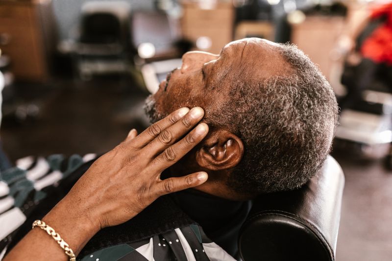
Why skin changes get missed on dark skin
Most scleroderma teaching images show light skin, and that costs Black patients diagnostic time. A 2024 systematic review of cutaneous systemic sclerosis in skin of color found that several classic skin clues, including telangiectasias (small dilated vessels), calcinosis, and visible digital swelling, were reported less often or were harder to spot on darker skin, which complicates recognition (Podwojniak, 2024).
What does show up, and is highly suggestive on dark skin, is salt-and-pepper pigmentation: patches where the skin loses pigment but keeps small pigmented spots around the hair follicles, giving a speckled look. It appears more often in patients with darker skin and can be an early tip-off. If a clinician dismisses skin tightening or pigment change as eczema or a tan line, ask directly whether scleroderma has been ruled out.
The dangerous organ complications
Three complications drive scleroderma deaths, and Black patients carry more of all three.
- Interstitial lung disease (ILD). Scarring of the lung tissue is the leading cause of scleroderma death. It often has no early symptoms, then shows up as breathlessness and a dry cough. Black race and anti-topoisomerase antibodies both track with more severe lung disease, so screening with lung function tests and a chest CT matters.
- Pulmonary hypertension. High pressure in the lung arteries strains the right side of the heart and causes breathlessness and fatigue. It is screened with an echocardiogram and confirmed with right heart catheterization.
- Scleroderma renal crisis. A sudden, dangerous spike in blood pressure with rapid kidney failure. It is a true emergency, more common in early diffuse disease and in people on higher-dose steroids, and it is treated with ACE inhibitor medication started fast.
How it is diagnosed
Diagnosis combines the exam with blood tests and a look at the nailfold. About 95% of people with systemic sclerosis have a positive antinuclear antibody (ANA) test, so that is usually the first screen. From there, three antibodies carry the most weight and appear in the formal ACR/EULAR classification criteria:
- Anti-topoisomerase I (anti-Scl-70) is linked to diffuse disease and interstitial lung disease, and it shows up more often in Black patients (31% versus 19% of white patients in the Johns Hopkins cohort).
- Anti-centromere is linked to the limited form and a generally slower course.
- Anti-RNA polymerase III is linked to diffuse disease and a higher risk of scleroderma renal crisis.
Nailfold capillaroscopy rounds it out. A clinician uses magnification to look at the tiny blood vessels at the base of your fingernails; enlarged or dropped-out capillaries are an early structural sign of systemic sclerosis and are part of the diagnostic criteria. Combined with the antibody pattern, this lets a rheumatologist separate scleroderma from other causes of Raynaud or skin tightening.
How it is managed
There is no cure that reverses the fibrosis, so management is organ-by-organ and aims to slow damage and protect function. Raynaud and digital ulcers are treated with vasodilator medications such as calcium channel blockers. Reflux is managed with proton pump inhibitors and meal-timing changes. Lung disease is treated with immunosuppressants such as mycophenolate and, in some cases, the antifibrotic nintedanib. Pulmonary hypertension has its own targeted drug classes. Scleroderma renal crisis is treated urgently with ACE inhibitors. Because the disease touches so many systems, care usually runs through a rheumatologist coordinating with pulmonology, cardiology, and nephrology. Early, consistent specialist follow-up is what bends the outcome curve, which is exactly the access gap that hits Black patients hardest.
How to get care
If you have Raynaud plus any skin tightening, puffy fingers, or new reflux, the move is a rheumatology referral and an ANA plus scleroderma antibody panel. Bring a written list of your symptoms and when they started, and ask specifically about lung screening, because early ILD is silent. If you want a clinician who understands how this disease presents on Black skin and takes your symptoms seriously, you can find a Black rheumatologist or Black-serving specialist in our directory. The patients who do best are the ones who get diagnosed and into specialist care before the lungs and kidneys are involved.
Frequently asked questions
Is scleroderma more common in Black people? ▼
Systemic sclerosis is not dramatically more common, but it is more severe in Black adults. Black patients develop it younger, are more likely to have the aggressive diffuse cutaneous form, have more interstitial lung disease, and have higher death rates than white patients in multiple cohort studies.
What is the first sign of scleroderma? ▼
For most people it is Raynaud phenomenon: fingers turning white, then blue, then red in cold or stress. Raynaud usually appears years before other signs. Puffy fingers, tightening skin on the fingers and face, and new acid reflux are the next common signals.
Can scleroderma skin changes look different on dark skin? ▼
Yes. Some classic clues like telangiectasias and visible swelling can be harder to spot on darker skin, which delays diagnosis. Salt-and-pepper pigmentation, where skin loses color but keeps pigment around hair follicles, is more visible on dark skin and can be an early tip-off.
What blood test detects scleroderma? ▼
There is no single test. Clinicians start with an ANA (positive in about 95% of patients), then check disease-specific antibodies: anti-Scl-70 (anti-topoisomerase, linked to diffuse disease and lung scarring), anti-centromere (limited disease), and anti-RNA polymerase III (diffuse disease and renal crisis risk). Nailfold capillaroscopy supports the diagnosis.
What is scleroderma renal crisis? ▼
A sudden, severe rise in blood pressure with rapid kidney failure. It is a medical emergency, more common in early diffuse disease and with higher steroid doses, and it is treated quickly with ACE inhibitor medication. A sudden severe headache, vision changes, or a blood pressure reading much higher than your normal warrants immediate care.