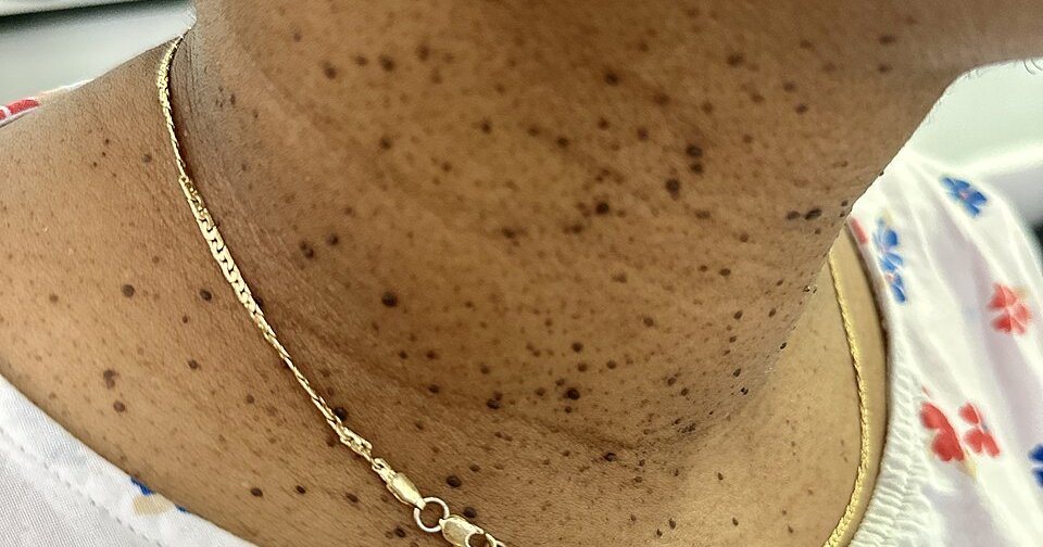
How to identify dermatosis papulosa nigra on Black skin
The bumps start earlier than most people remember. StatPearls describes lesions that first appear in adolescence as minute, round, skin-colored to dark brown macules resembling freckles, then become papular and grow in size and number with age. The mature lesion is a hyperpigmented, smooth-surfaced papule 1 to 5 mm across and 1 to 3 mm high, either flat-based or on a thin stalk.
Location is the strongest clue. They appear symmetrically across the malar cheeks, temples, and forehead, exactly the part of the face that catches the most light, and less often on the neck, upper chest, and back. Three negatives finish the identification: no scaling, crusting, or ulceration; no itch; and no spontaneous resolution, which is why the count climbs decade after decade instead of clearing up.
This is common, and the numbers back that up. Grimes and colleagues assessed 82 Black patients in a 1983 clinic study and found an overall incidence of 77 percent in that population, a female predominance of almost 2 to 1, and a peak count in patients in their sixties. The authors said their figure ran higher than average, and StatPearls puts reported incidences across selected study populations at 10 to 75 percent. StatPearls also says up to one third of African American adults in the United States are affected and reports positive family histories in 77 to 93 percent of cases. A 2020 Wake Forest survey of 50 African American adults, 48 of them women, found 86 percent had lesions on the face and 84 percent had a first-degree relative with the condition, at an average age at diagnosis of 44.7 years, decades after the bumps first show up.
The cause is not settled, and nothing you did brought them on. StatPearls says outright that the cause of dermatosis papulosa nigra is unknown. Hafner and colleagues found an activating FGFR3 mutation in both of the lesion samples they analyzed and none in the matched control tissue, the same gene mutated in seborrheic keratoses, and StatPearls says that shared genetic pathway may explain the predisposition that runs in families. Because the lesions sit in a photodistribution, StatPearls describes cumulative ultraviolet exposure as a proposed contributor and cites a finding that patients using topical skin-lightening products had worse involvement. Nothing about diet, hygiene, face oils, or touching your face causes this.
DPN, skin tags, and seborrheic keratoses: telling the three apart
Dermatosis papulosa nigra is the facial one. Sun-exposed skin, small uniform lesions, adolescent onset, strong family history, women about twice as often as men, and it typically occurs in Fitzpatrick skin types III through VI. StatPearls lists it as benign with no risk of malignant neoplasm and no link to any systemic disease.
Skin tags grow somewhere else and carry information about your health. The StatPearls chapter on acrochordons puts prevalence at roughly 50 to 60 percent of adults, affects men and women equally, and locates them in skin folds: neck, armpits, groin, and eyelids, hanging from a narrow stalk. The association is what matters. Skin tags are linked to obesity, type 2 diabetes, dyslipidemia, insulin resistance, and polycystic ovary syndrome, and StatPearls says multiple or recurrent lesions frequently signal metabolic derangements and warrant further evaluation. A neck full of skin tags is a reason to check your A1c and lipids. A face full of DPN is not.
Seborrheic keratoses are the larger, waxier growths the American Academy of Dermatology calls the barnacles of aging: growths that can get quite thick and take on a warty surface, arriving in middle age or later, ranging in color from white to black and turning up anywhere except the palms and soles. Most sources treat DPN as a variant of this one. StatPearls lays out the differences that keep the debate open: DPN stays on sun-exposed skin while seborrheic keratoses spread widely, DPN lesions are small and uniform while seborrheic keratoses vary widely in size, and DPN lacks the PIK3CA mutation found in seborrheic keratoses despite sharing the FGFR3 one. Metin and colleagues argue in Clinics in Dermatology that it is a distinct entity.
None of the three is a mole. A melanocytic nevus grows from pigment-producing cells, and none of these three does. When the appearance is genuinely unclear, a dermatoscope settles it without cutting: in Bhat's series of 100 patients the predominant pattern was fissures and ridges in a cerebriform arrangement, seen in 59 percent of cases, followed by comedo-like openings in 27 percent.
Removal is cosmetic, and the risk you are managing is pigment
Removal buys appearance, nothing else. StatPearls states that treatment is generally unnecessary and performed for cosmetic purposes, and that because darker skin types heal with dyspigmentation, all patients should be counseled that removal may cause it. That is the whole risk calculation on Black skin. In the 2010 randomized trial by Garcia, Azari, and Eisen, hyperpigmentation was the most common adverse outcome recorded. The 2025 systematic review by Jain, Caire, and Haas names postinflammatory hyperpigmentation a common adverse effect that often leads to dissatisfaction, and notes it can be mitigated with topicals that reduce local inflammation. Our guide to dark spots and hyperpigmentation on Black skin covers how those marks behave and how long they take to fade.
Common skin conditions are treated in primary care, and community health centers charge on a sliding scale that is often $0. Our directory of free and charitable clinics lists verified centers by state. A same-day telehealth visit is self-pay and cannot be billed to Medicaid or commercial insurance, so the clinic route usually costs less. If you need a prescription sooner than the next appointment:
Affiliate
StatPearls gives the rule that protects your whole face: initial treatment should be limited to a small number of lesions so postinflammatory dyspigmentation can be evaluated, and aggressive treatment avoided to minimize scarring and pigmentation problems. That is a test spot. Ask for one.
The techniques, and what the evidence says about each
Snip excision, light curettage, and light electrodesiccation are the standard three. StatPearls calls them the most common modalities, generally effective for smaller lesions, and specifies that electrodesiccation should be done at a low setting after topical anesthetic, with ointment on the crust daily until healed. Garcia and colleagues randomized 10 patients in an evaluator-blinded trial and measured mean clearance of 96 percent for curettage, 92.5 percent for electrodesiccation, and 88 percent for pulsed dye laser, with no significant difference between them. Half preferred electrodesiccation, and they rated the laser the most painful.
Lasers work, and the split-face trial is the one to know. Kundu and colleagues randomized 14 subjects with Fitzpatrick skin types IV through VI to two potassium-titanyl-phosphate laser treatments on one side of the face and two electrodesiccation treatments on the other. Blinded dermatologists found no significant difference between the sides, both were well tolerated, and the only thing separating them was comfort, which favored the laser. Tran and Richer catalog six laser devices reported to work, from the 532-nm KTP through the 10,600-nm carbon dioxide, and Maghfour and Ogunleye, reviewing 15 studies, found laser therapy increasingly used as safe and efficacious in skin of color. Ali and colleagues treated 45 patients with the carbon dioxide laser over a median of three sessions; of the 18 who answered the follow-up survey, 28 percent reported a few lesions coming back, and none reported scarring or a pigment change.
Low-intensity electrodesiccation has a large skin-of-color series behind it. Maruma, Dlova, and colleagues reviewed the records of 137 African patients, 72 percent of them Fitzpatrick type V. Adverse events were four cases of milia and two of scarring, and the authors concluded the technique was safe and cost-effective. They also flag the practical problem: laser devices carry high costs, which is why cheaper office techniques still matter.
Cryotherapy is the one to question. StatPearls says freezing is problematic because of the risk of hypopigmentation from melanocyte damage and should be used with extreme caution, and yet the Wake Forest survey found cryosurgery was one of the two methods patients most often received. If a clinician reaches for liquid nitrogen on your face, ask why that over electrodesiccation or a snip, and ask for a test spot first.
What removal looks like, from the chair to the mirror
Expect topical numbing cream, a short procedure, and small crusts where each lesion was. The crusts heal over the following days to a few weeks; the color takes longer. Aftercare is simpler than most people are told. Taylor, Averyhart, and Heath ran a double-blind, split-face pilot study in 20 African American subjects, treating wounds on one side of the face with a plain skin protectant ointment and the other with a topical antibiotic for 21 days. There were no differences in crusting, scabbing, healing, or postinflammatory hyperpigmentation at any time point. In that study, plain ointment did the job and the antibiotic added nothing.
Pigment settles last, and it can go either direction. The American Academy of Dermatology says that after removal of a seborrheic keratosis the skin may be lighter than the skin around it, that this usually fades with time, and that it is sometimes permanent. The Dakar series by Niang and colleagues evaluated 30 patients and removed lesions for those who wanted it; the result was satisfactory without scarring in 60 percent of the cases treated at day 45, with hypochromic scars persisting in four patients who practiced artificial depigmentation and one treated with carbon dioxide snow. Stop lightening products well before any procedure.
Removal is not a cure. StatPearls notes that lesions do not spontaneously resolve, that number and size increase with age and peak in the sixth decade, and that lesions may recur after any modality. Treat the face you have now and expect new bumps later. One thing to raise beforehand: if you have ever formed a keloid from a piercing, a cut, or surgery, say so. The published DPN treatment studies do not report keloids among their complications, but they were not designed to answer that question, and a keloid-prone history is worth putting on the table before anyone cuts or burns your face. Our guide to keloid scars on Black skin covers who tends to form them.
What it costs, and why insurance says no
Medicare's position is on the record. The local coverage determination titled Removal of Benign Skin Lesions, policy L35498, written by the contractor Wisconsin Physicians Service and revised effective for services on or after October 30, 2025, states that removal of certain benign skin lesions that does not pose a threat to health or function is considered cosmetic and is not covered by the Medicare program. Local determinations apply in the contractor's own jurisdictions, and other contractors publish their own versions of the same rule.
The same policy lists what does qualify as medically necessary: the lesion bleeds, itches, or causes pain; it changes in appearance, recently enlarges, or increases in number; it shows evidence of inflammation; it obstructs an orifice or restricts vision; there is clinical uncertainty where malignancy is a realistic consideration; a prior biopsy suggests malignancy; or the lesion sits where it takes repeated documented trauma. Read that list against the callout above on what is not DPN. The criteria are the same criteria.
The policy also spells out what happens when a patient wants an asymptomatic benign lesion removed anyway: the beneficiary becomes liable, and the physician must notify the patient in advance, with charges clearly stated. Commercial plans write their own benign-lesion policies, so ask yours by name. The 2025 systematic review is blunt about the consequence: management options are limited and often unaffordable, because treatment is elective in most cases. Ask the office for the price per session and the expected number of sessions in writing, and whether a test spot is billed separately.
When to see a dermatologist
Nothing here requires a visit. These earn one.
- Any bump that bleeds, crusts, ulcerates, itches, grows fast, or looks unlike the rest. That is the biopsy conversation, not the cosmetic one.
- You want them removed, with the technique chosen for your skin type rather than for whatever device is in the room.
- You have a history of keloids, or dark or light marks have lingered after a previous procedure.
- You use or recently used skin-lightening products, which the Dakar series tied to delayed healing and persistent hypochromic scars.
- The bumps are affecting how you move through the world. The Wake Forest survey measured an average Dermatology Life Quality Index score of six, a moderate effect on quality of life, and every patient there who had lesions removed reported their skin looked better. Only 36 percent had ever seen a physician about it.
Go in with three questions: is this dermatosis papulosa nigra or something that needs a biopsy, will you do a test spot before treating my whole face, and what is the total cost including follow-ups. You can find a Black dermatologist in our directory, along with clinicians who list a practice focus in skin of color.
Frequently asked questions
Are the small black bumps on my face moles? ▼
Almost certainly not. Moles come from pigment cells. Dermatosis papulosa nigra comes from the outer layer of skin, and StatPearls describes it as a benign epidermal growth with no risk of malignant neoplasm. The giveaways are pattern and family history: dozens of small, uniform, smooth bumps spread symmetrically across the cheeks, temples, and forehead, in someone whose parent or grandparent has them too.
What is the difference between dermatosis papulosa nigra and skin tags? ▼
Place and meaning. Skin tags grow in skin folds at the neck, armpits, groin, and eyelids, and affect men and women equally. StatPearls links them to obesity, type 2 diabetes, insulin resistance, and polycystic ovary syndrome, and says multiple lesions warrant a metabolic workup. Dermatosis papulosa nigra sits on sun-exposed facial skin, affects women about twice as often, starts in adolescence, and is tied to no systemic disease.
Can I remove dermatosis papulosa nigra at home? ▼
No. Every removal method leaves a small wound on facial skin that pigments readily, and StatPearls calls for low settings, topical anesthetic, and treating only a few lesions first to limit dyspigmentation. A home device or a drugstore acid does none of that, and you cannot tell from a mirror which bump needed a biopsy.
Does DPN removal leave dark marks on Black skin? ▼
It can, and that is the tradeoff. Hyperpigmentation was the most common adverse outcome in the 2010 randomized comparison of electrodesiccation, curettage, and pulsed dye laser, and the 2025 systematic review names it a common adverse effect that often drives dissatisfaction. The American Academy of Dermatology notes the opposite is also possible: skin can heal lighter than the area around it, usually fading with time and sometimes permanently. A test spot tells you which way your skin goes.
Will the bumps come back after removal? ▼
The treated ones usually stay gone, and new ones keep arriving. StatPearls reports that lesions do not spontaneously resolve, that number and size increase with age, and that patients should be counseled lesions may recur after any modality. In the largest reported carbon dioxide laser series, 28 percent of the patients who answered the follow-up survey reported a few returning.
Does insurance cover dermatosis papulosa nigra removal? ▼
Usually not. Medicare's local coverage determination on removal of benign skin lesions, policy L35498, states that removing certain benign lesions that do not threaten health or function is cosmetic and not covered, and that the beneficiary becomes liable for the cost. Coverage turns on symptoms: bleeding, itching, pain, recent change or enlargement, inflammation, obstruction, or genuine uncertainty about malignancy. Commercial plans write their own rules, so ask yours by name.