What tinea versicolor is, and why it turns Black skin lighter
Tinea versicolor, also called pityriasis versicolor, is an overgrowth of Malassezia, a lipid-loving yeast that lives on normal healthy skin. StatPearls describes it as a common, benign, superficial fungal infection, and notes that it is not a dermatophyte infection like the other conditions carrying the word tinea. Disease starts when the yeast shifts from its round yeast form into a filament form and spreads through the outermost layer of skin.
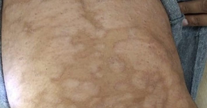
It lands where the oil glands are busiest: the upper back, the chest, the neck, the shoulders, and the upper arms. The patches are oval, well defined, and tend to run together into larger irregular areas over time. The face gets involved mainly in children.
On Black skin, the version most people meet is the light one. The 2014 International Journal of Dermatology review of tinea versicolor in dark-skinned individuals reports that most clinicians find the majority of dark-skinned patients present solely with hypopigmented lesions. The 2023 Life narrative review supplies the mechanism: Malassezia produces azelaic acid, which inhibits tyrosinase, the enzyme pigment cells need to build melanin. That review adds that the depigmenting effect is more visible in darker phototypes, and that hyperpigmented lesions are more common in lighter skin tones. StatPearls describes the same azelaic-acid mechanism and notes that affected skin stops darkening in response to sun, which is why the patches jump out in summer.
One study complicates that picture and belongs on the page. Aljabre and colleagues studied 100 cases in patients with skin types IV and V and concluded that in dark-skinned individuals tinea versicolor does not tend to be significantly hypopigmented. Both variants are real on Black skin. StatPearls notes that on darkly pigmented skin the hyperpigmented form runs from dark brown to grayish black. The rule that survives both findings: identify tinea versicolor by its shape, its location, and its scale, not by whether the patch is lighter or darker than the skin around it.
Risk factors here are environmental, not personal. StatPearls lists heat and humidity, oily skin, oily lotions and creams, pregnancy, malnutrition, oral contraceptives, and a weakened immune system, and reports that 21 percent of patients in one survey had a family history. The American Academy of Dermatology adds heavy sweating to that list, says people of all skin colors get it and that teens and young adults are most susceptible because their skin is oilier. Nothing on that list is a hygiene failure, and StatPearls says so directly: poor hygiene is not a causative factor.
What it gets mistaken for
Light patches on brown skin usually get one of two wrong answers, and the difference changes everything about what happens next.
Vitiligo. The American Academy of Dermatology names this confusion explicitly: when tinea versicolor causes light spots, it can be mistaken for vitiligo. The separation is clean. StatPearls describes vitiligo as completely depigmented macules and patches, and states that tinea versicolor only causes hypopigmentation. Vitiligo patches read chalk-white against surrounding skin, hold a sharp border, and carry no scale, and the 2023 Life review notes they often show pigment ringing the hair follicles and white hairs inside the patch. Vitiligo also favors the hands, the face, and the skin around the eyes and mouth rather than the oily upper back. If the patch is truly white rather than off-color, read our guide to vitiligo on Black skin and get evaluated, because vitiligo is an autoimmune condition with a different treatment path.
Ringworm. The shared word is misleading. Ringworm is tinea corporis, a dermatophyte infection, a different organism entirely from Malassezia, and StatPearls flags that distinction in its first paragraph. Ringworm makes a ring with a raised, actively scaling edge and a clearer center, and it usually itches. Tinea versicolor makes flat oval patches in clusters on the trunk that are asymptomatic or mildly itchy. Some antifungals overlap, but the diagnosis, the course, and the recurrence pattern do not. Our guide to ringworm on Black skin covers that one.
Pityriasis alba. A mild eczematous dermatitis, per StatPearls, appearing as hypopigmented macules and small patches on the face and, less often, the upper arms, mostly in children with a history of atopic dermatitis. Its borders are poorly defined where tinea versicolor's are sharp.
Post-inflammatory hypopigmentation. Light marks left behind by acne, eczema, a burn, or a procedure. A 2023 Journal of Clinical Medicine review describes it as a common acquired pigmentary disorder that is more prominent in skin of color and one that mostly resolves on its own over time. It traces the footprint of whatever rash came before it, and it does not scale. The mirror-image problem, dark marks instead of light ones, is covered in our guide to dark spots and hyperpigmentation on Black skin.
Hypopigmented mycosis fungoides. This is the one worth knowing about. It is a cutaneous T-cell lymphoma, and StatPearls notes that the hypopigmented variant is most prevalent among people with dark complexions, showing up as light patches on the trunk and extremities. It does not clear with antifungals. Light patches that survive a full course of correct treatment and keep spreading need a biopsy, not a second round of shampoo.
Seborrheic dermatitis, secondary syphilis, erythrasma, guttate psoriasis, and confluent and reticulated papillomatosis fill out the differential in StatPearls. The 2014 review of dark-skinned patients lists confluent and reticulated papillomatosis, seborrheic dermatitis, pityriasis rosea, pityriasis alba, and vitiligo.
Common skin conditions are treated in primary care, and community health centers charge on a sliding scale that is often $0. Our directory of free and charitable clinics lists verified centers by state. A same-day telehealth visit is self-pay and cannot be billed to Medicaid or commercial insurance, so the clinic route usually costs less. If you need a prescription sooner than the next appointment:
Affiliate
How the diagnosis gets confirmed
Most of the time a dermatologist recognizes it on sight. When there is doubt, two tools settle it, and one of them is weaker than its reputation.
A KOH scraping is definitive. Scale is scraped onto a slide, soaked in potassium hydroxide, and read under a microscope. StatPearls describes the classic finding as short hyphae plus grape-like clusters of yeast cells, a pattern often likened to spaghetti and meatballs. No biopsy is required.
A Wood's lamp misses more than half of cases. Affected skin can fluoresce gold-yellow, yellow-green, or coppery-orange under ultraviolet light, and the American Academy of Dermatology describes the device held four or five inches from the skin. StatPearls reports that fluorescence is present in fewer than 50 percent of affected patients. A dark reading does not rule tinea versicolor out. If a clinician waves a lamp, sees nothing, and moves on, ask for the scraping.
The treatment ladder, from drugstore to prescription
Topical medication is first-line at every level of severity short of extensive disease. StatPearls is explicit that systemic therapy is not the opening move for limited tinea versicolor.
Start over the counter for mild, limited patches. The American Academy of Dermatology lists shampoo containing selenium sulfide, and creams or ointments containing miconazole, clotrimazole, or terbinafine, as products available without a prescription. Their instructions: wash and dry the affected skin, apply a thin layer of cream once or twice a day for at least two weeks, and when using a shampoo leave it on 5 to 10 minutes before rinsing. Treat the whole patch plus a margin of normal-looking skin around it, since the organism does not respect the visible border.
Prescription topicals are stronger versions of the same idea. StatPearls lists ketoconazole 2 percent shampoo applied for five minutes and rinsed, used over one to three days, with three days preferred given how often the condition returns. In a randomized study it cites, roughly 80 percent of patients reached mycologic cure with either a single application or three consecutive days. Its other listed options: selenium sulfide 2.25 to 2.5 percent applied for 10 minutes daily for one week, topical terbinafine 1 percent once or twice daily for one to four weeks, ciclopirox 1 percent twice daily for two weeks, and zinc pyrithione 1 percent for five minutes daily for two weeks.
Pills are for widespread or treatment-resistant disease. StatPearls names itraconazole 200 mg daily for seven days and fluconazole 300 mg weekly for two weeks as the preferred oral agents. A 2014 systematic review in the Journal of Cutaneous Medicine and Surgery pooled 57 trials and landed on the same regimens: itraconazole 200 mg per day for five or seven days, and fluconazole 300 mg per week for two weeks. These are monitored prescriptions, because systemic azoles can disturb liver function tests and interact with other medications.
Two oral drugs that do not work here. Oral terbinafine is ineffective against tinea versicolor even though the cream form works, per StatPearls and the 2015 Journal of Fungi review by Gupta and Foley. StatPearls adds that griseofulvin is ineffective here too. Terbinafine pills are the standard for toenail and scalp fungus, which makes this a common wrong turn. Oral ketoconazole is off the table entirely: StatPearls cites life-threatening liver toxicity, adrenal insufficiency, and drug interactions, and Gupta and Foley write that it should no longer be prescribed.
After treatment: the pigment timeline nobody explains
This is where most people decide the treatment failed, and most of them are wrong.
The infection and the color run on two different clocks. The 2023 Life review states that the disappearance of scale confirms therapeutic success, and that further weeks to months may be needed for complete repigmentation. StatPearls says restoration of normal pigmentation may take months following treatment, and that pigment changes most of the time resolve within 2 to 4 months of starting treatment. The American Academy of Dermatology puts it in patient language: with treatment the yeast is easy to kill, but the skin may stay lighter or darker for weeks or months before it returns to its normal color.
StatPearls names the consequence outright. Hypopigmentation and hyperpigmentation persisting for months after successful treatment lead patients to inaccurately believe the treatment was unsuccessful. The same page gives clinicians the instruction that reader and doctor both need: confirm treatment failure before retreating, because pigment change outlives the fungus. Active infection means scale and a positive KOH prep. A smooth, pale, scale-free patch is healing skin.
Two things speed the visible recovery. Daily broad-spectrum sunscreen keeps the surrounding skin from tanning away from the patch, which shrinks the contrast while the pigment cells restart. And time, which is not a satisfying answer but is the accurate one.
Recurrence is the rule, and maintenance is the answer
The 2023 Life review puts the recurrence rate as high as 80 percent. That is not a statistic about bad treatment, it is a consequence of biology. Gupta and Foley explain that the odds of relapse are high precisely because Malassezia is a permanent resident of normal skin flora. No course of medicine evicts it. Treatment returns an overgrowth to baseline, and heat, humidity, and sweat push it forward again.
So the plan after clearance is a maintenance plan. StatPearls describes prophylaxis with topical selenium sulfide 2.5 percent or ketoconazole 2 percent shampoo applied to the entire body for ten minutes once per month. The American Academy of Dermatology recommends a medicated cleanser once or twice a month, especially during warm and humid periods, and notes that people in tropical climates may need it year round. For people who relapse constantly, StatPearls cites oral itraconazole prophylaxis, 200 mg twice in one day, once a month. In the trial behind that number, as Gupta and Foley describe it, 88 percent of patients on monthly itraconazole were still mycologically clear at six months versus 57 percent on placebo.
The honest limit on that advice: Gupta and Foley note that research evaluating prophylactic antifungal treatment is limited, and the 2023 Life review says the whole-body antifungal wash approach is still poorly supported in the literature. It is cheap, it is low-risk, and the clinical bodies recommend it, and the evidence behind the monthly wash is thinner than the evidence behind the initial treatment. Both facts belong in the decision.
When to see a dermatologist
Over-the-counter treatment handles mild, limited tinea versicolor. Book a visit when:
- A full two-week course of an over-the-counter antifungal has not cleared the scale.
- The patches cover a large share of the trunk, or sit somewhere you cannot reach to treat.
- It has returned more than twice in a year and you want a maintenance regimen instead of another round of guessing.
- The patches never scale, never respond, and keep spreading, which is the pattern that warrants a biopsy to rule out hypopigmented mycosis fungoides.
- Patches are turning chalk-white, moving to the hands or the face, or growing white hairs inside them, which points toward vitiligo instead.
- You are immunosuppressed, living with poorly controlled diabetes, or on long-term steroids. StatPearls flags recurrence, drug resistance, and dissemination as reasons to consider an immunodeficient state.
Bring the pigment question into the room and say it plainly, because the discoloration is the part that affects how you live. The 2023 Life review documents that skin discoloration is a major concern for these patients, especially in darker skin types, where it is more common and more apparent. Ask two questions: is there still active infection here, and what is my monthly maintenance plan. You can find a Black dermatologist in our directory, along with clinicians who list a practice focus in skin of color.
Frequently asked questions
Is tinea versicolor contagious? ▼
No. The yeast that causes it, Malassezia, is already part of normal skin flora on healthy people. StatPearls states plainly that tinea versicolor is not contagious and that poor hygiene is not a causative factor. You did not catch it from anyone and you cannot give it to your partner, your kids, or a teammate.
Why are my patches lighter than the rest of my skin? ▼
Malassezia produces azelaic acid, which blocks tyrosinase, the enzyme pigment cells use to make melanin. The 2023 Life review notes that this depigmenting effect is more visible in darker phototypes, while hyperpigmented lesions are more common in lighter skin tones. The affected skin also stops tanning, so the contrast gets sharper in summer.
How do I know if it is tinea versicolor or vitiligo? ▼
Scale and shade. Vitiligo is complete loss of pigment: chalk-white, sharply bordered, with no scale, sometimes with white hairs growing inside the patch. Tinea versicolor only lightens skin, it never depigments it, and scratching the patch raises a fine powdery scale. A dermatologist settles it in minutes with a scraping under the microscope. Our guide to vitiligo on Black skin covers that condition in full.
How long does it take for the white patches to go away after treatment? ▼
Weeks to months. The infection clears fast, but repigmentation runs on a separate clock. StatPearls reports that pigment changes usually resolve within 2 to 4 months of starting treatment, and the American Academy of Dermatology says skin may stay lighter or darker for weeks or months before returning to its normal color. A pale patch with no scale is healing skin, not active infection.
Why does tinea versicolor keep coming back every summer? ▼
Because treatment knocks back an overgrowth of an organism that lives on you permanently. Heat, humidity, and sweat push it into its filament form again. The 2023 Life review puts the recurrence rate as high as 80 percent. That is why the American Academy of Dermatology recommends a medicated cleanser once or twice a month during warm and humid periods instead of waiting for the patches to return.
Do over-the-counter antifungal shampoos actually work? ▼
For mild, limited disease, yes. The American Academy of Dermatology lists shampoo containing selenium sulfide, and creams containing miconazole, clotrimazole, or terbinafine, as products you can buy without a prescription, applied once or twice a day for at least two weeks, with shampoo left on 5 to 10 minutes before rinsing. If a full course does not clear the scale, that is the point to see a dermatologist for prescription strength or oral therapy.