If you have a round patch on your scalp, ear or cheek with a dark rim and a lighter center, or a smooth bald spot where hair used to grow, ask a dermatologist for a biopsy now, not after it spreads. That pattern can be discoid lupus erythematosus (DLE), and on the scalp it destroys hair follicles for good once scar tissue forms. In a Dallas registry of 112 Black patients with DLE, 87% had it on the scalp and 79% had scarring hair loss.
What discoid lupus looks like on brown and black skin
DLE is the most common chronic form of cutaneous lupus, the type that lives in the skin. Common sites are the scalp, ears and cheeks, and it can also appear on the face, the V of the neck and the backs of the arms. A 2023 review in Anais Brasileiros de Dermatologia describes how a lesion changes over time: it starts as a firm, raised plaque, develops scale plugged into the hair follicles, then thins and changes color, with a lighter center and a darker edge.
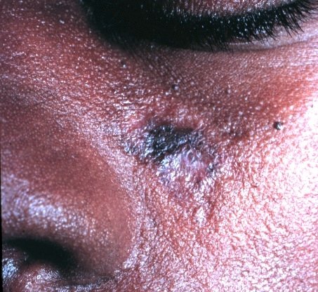
On melanin-rich skin, that color change is the feature you are most likely to notice. In the Dallas registry (UT Southwestern, 2009 to 2020), 111 of 112 Black patients had dyspigmentation, and 59% had lesions on their ears. The researchers note that a pale center stands out more against darker surrounding skin, and the dark rim can be more pronounced. A 2024 case report in JAAD Case Reports adds that in darker skin tones DLE can leave fully depigmented bald areas.
Expert criteria list the active plaque as red to violet, with follicular plugs, thinned scarred skin and color change. Scalp patches can itch, burn or feel tender, and some cause no symptoms at all, according to a review of hair loss in lupus. Friction and injury can trigger new spots: a 2025 case described an African American woman whose plaques formed exactly where her eyeglass frames pressed on her face.
On the scalp, scarring hair loss can be permanent
The scalp is the most common site of DLE, involved in 30% to 50% of reported cases. Early lesions are only mildly inflamed and have not scarred yet. Even so, as many as 60% of people with scalp DLE progress to permanent hair loss, and once fibrous tissue replaces the follicle, the hair does not come back (Concha and Werth, 2018). In the Dallas registry, 82% of Black patients had scalp color change and 79% had scarring alopecia.
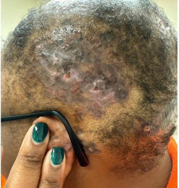
That is why timing matters. As DLE progresses, patches become thin, pale and empty of follicle openings, a 2024 case report notes (JAAD Case Reports). Treatment at that point protects the hair around the scar. Caught earlier, hair can return: a 2024 case documented full regrowth of a scalp DLE patch a year after hydroxychloroquine and pimecrolimus cream.
Other scarring hair losses on textured hair, including central centrifugal cicatricial alopecia (CCCA) and long-standing traction alopecia, can look similar. One clue: in scalp DLE, redness, scale and color change are most prominent in the center of the bald patch, while in other scarring alopecias activity tends to sit at the rim. One reported African American patient was treated for DLE until a biopsy showed a different blistering disease entirely (Concha and Werth). The Dallas researchers recommend gentle hair care for Black patients with scalp DLE to limit further damage.
An over-the-counter minoxidil from any pharmacy is the same active ingredient a paid prescription service sells at a markup, and a community health center can check for an underlying cause on a sliding scale. Our directory of free and charitable clinics lists centers by state. A same-day telehealth visit is self-pay and is not billed to Medicaid, so try the pharmacy shelf and a clinic workup first. If you want a telehealth visit instead:
Talk to a clinician about hair loss.
- Online consult on causes and prescription options
- Price shown before you book
 with Sesame Care
Affiliate link, we may earn a commission at no extra cost to you.
Get a punch biopsy from a dermatologist
Diagnosis is a skin biopsy, which a 2024 case report calls the gold standard. For scalp lesions, the North American Hair Research Society recommends at least one 4 mm punch biopsy taken from an area of active hair loss, deep enough to include the fat beneath the skin (Concha and Werth). Ask that the sample come from an active area rather than old scar.
The Dallas researchers name a real barrier: some Black patients report that clinicians seem unfamiliar or uncomfortable with Black skin and hair. They recommend that Black patients with suspected DLE have both scalp and ears examined. Many dermatologists also use a dermatoscope, a handheld magnifier that shows plugged follicles. You can search for one by city in our dermatology directory.
Up to 28% of people with discoid lupus develop systemic lupus
DLE can stay in the skin, or it can be the first sign of systemic lupus erythematosus (SLE), which can involve joints, blood and organs. A 2012 review in the British Journal of Dermatology puts the share who develop SLE at up to 28%. The shift can come fast: among 32 patients at an academic lupus center who progressed, the median time from DLE to SLE was 453 days.
The same review lists warning signs that travel with progression: widespread skin lesions, joint pain or swelling, nail changes, anemia, a low white cell count, a high sedimentation rate and high ANA levels. It recommends full skin exams, joint checks and labs including ANA, ESR and a complete blood count at regular visits. If you have DLE and develop joint pain or swelling, tell your doctor, and read our guide to lupus symptoms and diagnosis in Black women.
Treatment starts with sun protection and topical steroids
Ultraviolet light triggers lesions in many people with cutaneous lupus. In a randomized trial of 25 patients, no participant developed UV-induced lupus lesions on sunscreen-treated skin. Daily broad-spectrum sunscreen, a hat for scalp disease and long sleeves are the baseline.
For active plaques, the usual start is a potent steroid cream or ointment, used for the shortest effective time. A Cochrane review found five small trials of drugs for DLE, none of high quality; in one, lesions cleared completely in 27% of people using fluocinonide cream and 10% using hydrocortisone. Thick, stubborn plaques can be treated with steroid injections into the lesion, and tacrolimus or pimecrolimus can replace steroids when treatment has to continue for months (review).
Hydroxychloroquine needs eye screening from the start
When creams are not enough or lesions are widespread, antimalarial pills are the first-line systemic treatment, and hydroxychloroquine is the one most used. The drug can damage the retina over years of use, and the damage is not reversible. The American Academy of Ophthalmology's 2025 revision of its screening recommendations sets these rules:
- A daily dose of no more than 5 mg per kilogram of real body weight.
- A baseline exam with retinal imaging (OCT and fundus autofluorescence) soon after you start.
- Yearly OCT and autofluorescence screening while you take it, which can be deferred during the first 5 years if you have no significant risk factors.
- Higher risk with kidney disease, tamoxifen use, or starting the drug at an older age.
Bring your weight and dose to your eye appointment, and ask the ophthalmologist to send results to your dermatologist.
Smoking cuts the odds that treatment works
A meta-analysis of 10 studies and 1,398 patients found smoking linked to a 2-fold drop in the proportion of people with cutaneous lupus who improved on antimalarials. In a cohort of 218 patients, current smokers had worse disease and worse quality of life, and smoking has been found to worsen scalp DLE and make it harder to treat. Quitting is part of treatment.
One more reason to keep follow-up visits: long-standing DLE sores that ulcerate or bleed should be checked for squamous cell skin cancer (Concha and Werth). Any old patch that changes, opens or will not heal deserves a look.
Frequently asked questions
What does discoid lupus look like on Black skin? ▼
Round or coin-shaped plaques, often on the scalp, ears or cheeks, that start firm and scaly and heal with a lighter center and a darker rim. In one registry, 111 of 112 Black patients with DLE had color change. Fully depigmented patches can appear in darker skin.
Does hair grow back after discoid lupus on the scalp? ▼
It can while the patch is still active and the follicles are intact. Once scar tissue replaces the follicle, the hair loss is permanent. That is why early biopsy and treatment matter most on the scalp.
Can discoid lupus turn into systemic lupus? ▼
In some people. Up to 28% of people with DLE develop systemic lupus, and in one cohort the median time to progression among those who did was 453 days. Regular ANA tests, blood counts and joint checks are how doctors watch for it.
How is discoid lupus different from CCCA? ▼
Both cause scarring hair loss on textured hair. In scalp DLE, redness, scale and color change are most prominent in the center of the bald patch, and DLE often appears on the ears and face too. A punch biopsy from an active area tells them apart.
Do I need eye exams if I take hydroxychloroquine for discoid lupus? ▼
Yes. The American Academy of Ophthalmology advises a baseline exam soon after starting, then yearly retinal screening with OCT and autofluorescence. Yearly screening can be deferred for the first 5 years if you have no significant risk factors.